44 human heart diagram with labels
Human heart: Anatomy, function & facts | Live Science The human heart is an organ that pumps blood throughout the body via the vessels of the circulatory system, supplying oxygen and nutrients to the tissues and removing carbon dioxide and other ... Male Reproductive System: Labeled Diagram of Organs - Study.com Penis. The penis is the organ directly involved in sexual intercourse. It is composed of 3 parts: the root, body, and glans. The root is directly attached to the abdominal wall.
Anatomy, Thorax, Heart - StatPearls - NCBI Bookshelf The heart is a muscular organ situated in the center of the chest behind the sternum. It consists of four chambers: the two upper chambers are called the right and left atria, and the two lower chambers are called the right and left ventricles. The right atrium and ventricle together are often called the right heart, and the left atrium and left ventricle together functionally form the left ...
Human heart diagram with labels
Diagrams, quizzes and worksheets of the heart | Kenhub Worksheet showing unlabelled heart diagrams. Using our unlabeled heart diagrams, you can challenge yourself to identify the individual parts of the heart as indicated by the arrows and fill-in-the-blank spaces. This exercise will help you to identify your weak spots, so you'll know which heart structures you need to spend more time studying ... Heart histology: Cells and layers - Kenhub Gross anatomy Location. The heart is a muscular, four-chambered system that is responsible for pumping blood through the vascular network. The organ is located within the thoracic cavity in a region known as the mediastinum.It is bordered bilaterally by the lungs, anteriorly by the sternum and posteriorly by the oesophagus and thoracic vertebra. Anatomy of The Human Ribs - With Full Gallery Pictures! The Anatomy of the Human Ribs (costae) are one of the integral parts of the chest wall; they make up the lateral part of our body, its anterior and posterior wall and they entirely build the lateral parts of the chest wall.. The anatomy of the human ribs is made up of 24 ribs. These ribs are parted in 12 pairs (each on the left and right side of the chest wall), with the sternum, metasternum ...
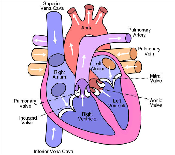
Human heart diagram with labels. Identify Various Parts Of A Human Heart: Trivia Quiz - ProProfs The heart is the most important organ in the body. It is in charge of keeping the processes within the body moving by facilitating the transfer of blood throughout the body. The quiz below is to test out interesting facts you may know about the heart. Give it a try and good luck. › photos › muscular-systemMuscular System Labeled Diagram Stock Photos, Pictures ... Labeled Muscles of the Human Body, Anterior View, 3D Rendering Frontal view of the muscular system of the male human body with descriptive labels pointing to the muscles on a white background. muscular system labeled diagram stock pictures, royalty-free photos & images How the Heart Works: Diagram, Anatomy, Blood Flow The heart is an amazing organ. It starts beating about 22 days after conception and continuously pumps oxygenated red blood cells and nutrient-rich blood and other compounds like platelets throughout your body to sustain the life of your organs.; Its pumping power also pushes blood through organs like the lungs to remove waste products like CO2.; This fist-sized powerhouse beats (expands and ... Histology, Heart - StatPearls - NCBI Bookshelf The heart is a four-chambered organ responsible for pumping throughout the body. It receives deoxygenated blood from the body, sends it to the lung, receives oxygenated blood from the lungs, and then distributes the oxygenated blood throughout the body. At the histological level, the cellular features of the heart play a vital role in the normal function and adaptations of the heart.
Heart - Wikipedia The human heart is situated in the mediastinum, at the level of thoracic vertebrae T5-T8.A double-membraned sac called the pericardium surrounds the heart and attaches to the mediastinum. The back surface of the heart lies near the vertebral column, and the front surface sits behind the sternum and rib cartilages. The upper part of the heart is the attachment point for several large blood ... en.wikipedia.org › wiki › File:Diagram_of_the_humanFile:Diagram of the human heart (cropped).svg - Wikipedia Diagram of the human heart, created by Wapcaplet in Sodipodi. ... Add Inferior vena cava and pericardium labels: 18:08, 14 August 2018: 656 × 631 (209 KB) Jmarchn: Diagram of Human Heart and Blood Circulation in It Four Chambers of the Heart and Blood Circulation. The shape of the human heart is like an upside-down pear, weighing between 7-15 ounces, and is little larger than the size of the fist. It is located between the lungs, in the middle of the chest, behind and slightly to the left of the breast bone. The heart, one of the most significant organs ... How the Heart Works - The Heart | NHLBI, NIH Blood also carries carbon dioxide to your lungs so you can breathe it out. Inside your heart, valves keep blood flowing in the right direction. Your heart's electrical system controls the rate and rhythm of your heartbeat. A healthy heart supplies your body with the right amount of blood at the rate needed to work well.
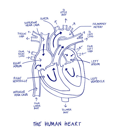
Heart to Heart - Lesson - TeachEngineering After Part II of this lesson, students should be able to: Identify the parts of the human heart on a diagram and with a biological specimen. Describe blood flow through the human heart, elaborating on what role each part of the heart plays in this process. Define terms associated with the heart and its function. Labeled diagram of the heart along with the various ... - Kelsi Schanbacher Blood circulation, heart chambers, coronary arteries and other components of a healthy heart from froedtert. Labeled diagram of the heart along with the various blood vessels and valves. Beating over 35 million times per year or 100,000 per day, . Grade 9 worksheet 2 on circulatory . Heart diagram of coronary artery function . The Heart's Electrical System: Anatomy and Function This pushes their load of blood into the right and left ventricles, the bottom two chambers of the heart. 1. The electrical signal then passes through the AV node to the ventricles, where it causes the ventricles to contract in turn. The cardiac electrical signal controls the heartbeat in two ways. First, since each electrical impulse generates ... › 1-label-the-heartLabel the heart — Science Learning Hub Jun 16, 2017 · In this interactive, you can label parts of the human heart. Drag and drop the text labels onto the boxes next to the heart diagram. If you want to redo an answer, click on the box and the answer will go back to the top so you can move it to another box.
Heart Diagram – 15+ Free Printable Word, Excel, EPS, PSD Template Download | Free & Premium ...
Circulatory System Diagram - New Health Advisor Heart: Human heart is a powerful pumping organ that pushes out the oxygenated blood from the left ventricle to be distributed to the periphery; whereas right heart collects deoxygenated blood ... There are different types of circulatory system diagrams; some have labels while others don't. The color blue stands for deoxygenated blood while ...

Unlabelled Diagram Of The Heart# - ClipArt Best
Human Heart for Kids: 2 Fun Heart Models plus Worksheets We also make a playdough heart model and completed the free printable heart worksheets. This is just one of our human body activities as part of our human body for kids unit studying anatomy for grade 1, grade 2, grade 33, grade 4, grade 5, grade 6, and garde 7 students. Whether you are a parent, teacher, or homeschooler you will love these ...

Heart Diagram Unlabeled | Medical anatomy, Medical education, Medical school studying
› photos › diagram-of-bodyDiagram Of Body Organs Female Pics Pictures, Images ... - iStock Heart disease and atherosclerosis prevention infographics. Healthy lifestyle concept. Heart disease and atherosclerosis prevention infographics. Healthy lifestyle concept. Vector flat illustration. Prevention cardiovascular problem. Weight scale, heart, exercise, food, diabetes control diagram of body organs female pics stock illustrations

Human Heart Diagram - Unlabeled - Tim's Printables
Heart: illustrated anatomy - e-Anatomy - IMAIOS This interactive atlas of human heart anatomy is based on medical illustrations and cadaver photography. The user can show or hide the anatomical labels which provide a useful tool to create illustrations perfectly adapted for teaching. Anatomy of the heart: anatomical illustrations and structures, 3D model and photographs of dissection.
Human Heart Anatomy Diagram Blue Line On A White Background Stock Illustration - Download Image ...
Heart Drawing With Label : Human Heart Diagram Images Stock Photos ... Most frequent question in exam to draw human heart diagram with labels. Png, jpg, gif · no labels version · azərbaycanca · català · english · english · hrvatski · italiano · lingua franca nova. Internal structure of human heart shows four chambers viz. Cell structure and functions / animal cell vs plant cell / parts of cell / ch 8 ...
Anatomy Review: The Heart
Body Cavities and Membranes: Labeled Diagram, Definitions Body cavity labeled diagram of organs they contain, membranes, and lateral views. Body cavity definitions and subdivisions in tables and charts. Ventral, dorsal, cranial, spinal, vertebral, thoracic, pleural, pericardial, mediastinum, abdominopelvic, abdominal, and pelvic cavities explained. Quiz yourself with the labeled views!

Dentistry and Medicine: Thorax,Lungs,Heart Anatomy and Physiology Diagrams Free Download
Basic Human Heart Diagram Labeled - Simple Human Lungs And Heart ... Labeled human heart fileheart diagram ensvg wikipedia. This science quiz game will help you identify the parts of the human heart with ease. Human heart beats over 100,000 times a day? These are the two upper chambers, which receive blood. Drag and drop the labels to identify the different parts of the human heart.

Sketch Human Heart Labelled Diagram - Aflam-Neeeak
20 Free Printable Heart Templates, Patterns & Stencils The three heart shapes are: classic heart, tapered heart, and rounded heart. The heart templates come in varying sizes, from large (approximately 7 inch size, 1 per page), medium (5 inch, 2 per page), small (3 inch, 6 per page), and mini (2 inch, 15 per page). There's also a page of mixed shapes and sizes from large to mini hearts.

The Heart Diagrams Labeled and Unlabeled
byjus.com › biology › diagram-of-heartHeart Diagram with Labels and Detailed Explanation - BYJUS The human heart is the most crucial organ of the human body. It pumps blood from the heart to different parts of the body and back to the heart. The most common heart attack symptoms or warning signs are chest pain, breathlessness, nausea, sweating etc. The diagram of heart is beneficial for Class 10 and 12 and is frequently asked in the ...

The Heart and Circulatory System | Teaching Resources
Human Circulatory System | Parts, Functions, Types Human Heart with parts label. The circulatory system is a network that carries blood throughout the body. The human circulatory system supplies the food and oxygen that is necessary for the body to survive. At the same time, it carries carbon dioxide and other waste materials away from ...

Human&Animal Anatomy and Physiology Diagrams: Heart and Great Vessels Diagram
Free Circulatory System Worksheets and Printables This will help your children memorize the different parts of the circulatory system by using the free labels included. Inside Out Anatomy Circulation Worksheet - This worksheet gives you a look into pulmonary circulation and the path of oxygen-rich blood through the heart. Awesome Anatomy Heart Diagram Worksheet - The Awesome circulatory ...

31 Blood Vessels Diagram To Label
Mnemonics for Heart Anatomy and Physiology (Video) - Mometrix A. Murmurs indicate abnormal or turbulent blood flow through specific vessels or chambers of the heart. The most common causes of heart murmurs can be remembered with the mnemonic SPAMS, S tenosis of a valve, P artial obstruction, A neurysm, M itral or aortic regurgitation, and S eptal defect. Q.
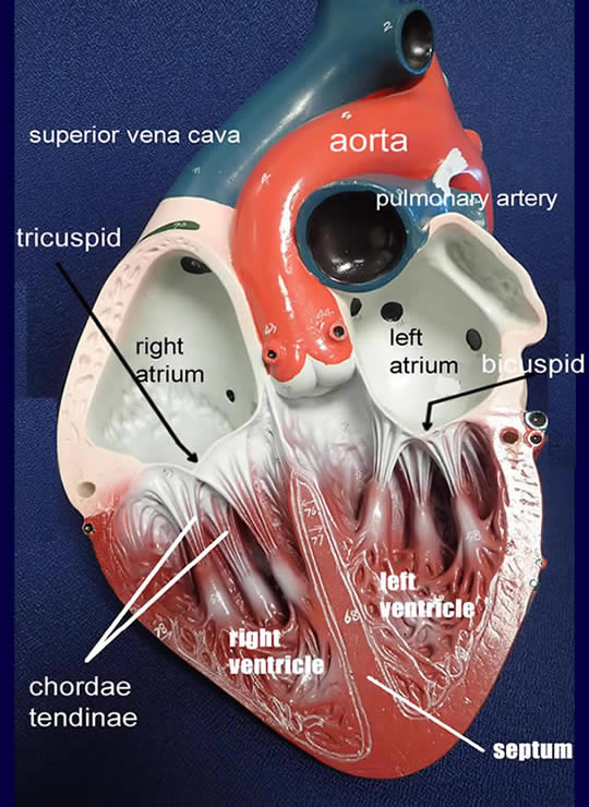
Heart Models
Heart Labeling Quiz: How Much You Know About Heart Labeling? Here is a Heart labeling quiz for you. The human heart is a vital organ for every human. The more healthy your heart is, the longer the chances you have of surviving, so you better take care of it. Take the following quiz to know how much you know about your heart. 1.

Heart diagram, Human heart anatomy, Human heart diagram
Free Heart Worksheets for Human Anatomy Lessons These human heart worksheets are a fun way to teach your kids about the human heart. Among other activities, your kids can label the parts of the heart, work with Montessori 3-part cards, and color. The Heart Nomenclature Cards - This paid resource is formatted as the Montessori 3-part cards. The cards will help your kids learn 23 parts of ...

Label Kidney Diagram - Human Anatomy
› photos › anatomy-of-the-human-body29,047 Anatomy Of The Human Body Premium High Res Photos Browse 29,047 anatomy of the human body stock photos and images available, or start a new search to explore more stock photos and images.
Respiratory System Worksheet - WikiEducator
Anatomy of the heart and coronary arteries (coronary CT) - IMAIOS Anatomy of the human heart and coronaries: how to view anatomical structures. This tool provides access to an MDCT atlas in the 4 usual planes, allowing the user to interactively discover the heart anatomy. The images are labeled, providing an important medical and anatomical tool. The quiz mode makes it possible to evaluate the user's progress.
![BIOMED ALL INVITED: The Human Heart [ANATOMY/PHYSIOLOGY/CONDUCTION SYSTEM]](https://blogger.googleusercontent.com/img/b/R29vZ2xl/AVvXsEiQ-LhlyIFCsOGX3N-6vD9of9pmpcvEnoWh5R8gre22MhyphenhyphenIEK1Nc2PFMWwguq_lh1bAxvz6KA7n_xeT8Xa1UkIRrPRjGx1zCY-4AH6H-b_CNVyXoVdQb6Ctu-KY4xrJVGlyuXx61ZbJWNBi/s1600/heart+anatomy.jpg)
BIOMED ALL INVITED: The Human Heart [ANATOMY/PHYSIOLOGY/CONDUCTION SYSTEM]
Heart & Circulatory System Diagram, Parts & Function, For Kids The circulatory system includes the heart and blood vessels. The blood vessels distribute blood, which delivers oxygen and other nutrients to the cells. It also helps eliminate wastes, such as transport hormones and carbon dioxide, and maintains the body's fluid balance and temperature (1). Blood comprises blood cells (red and white) and plasma.
Post a Comment for "44 human heart diagram with labels"