45 microscope images with labels
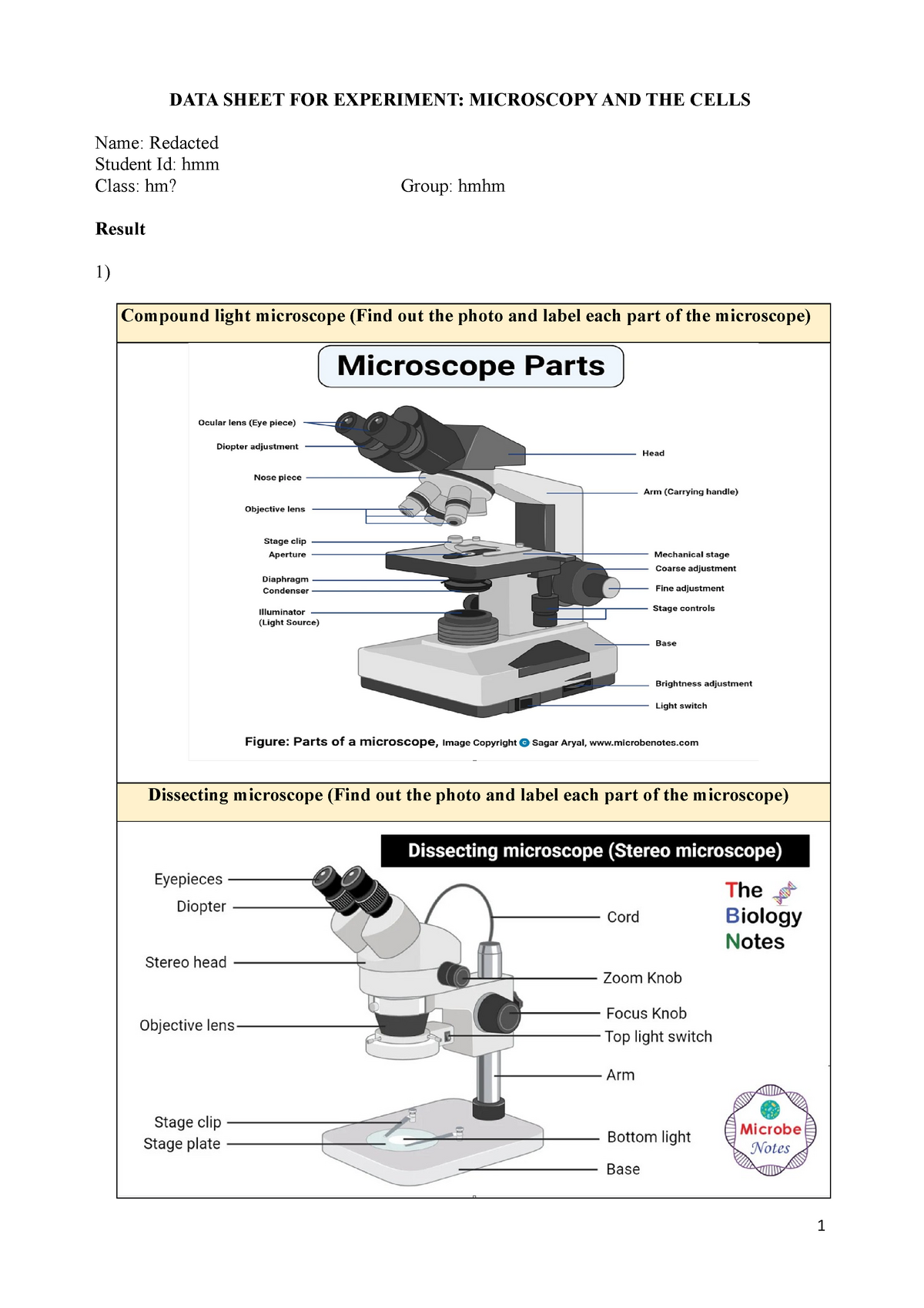
Mitosis Images Labeled | Virtual Anatomy Lab VAL - ncccval Endocrine Rabbit Dissection Unlabeled. Cardiovascular. Cardiovascular Histology Labeled. Cardiovascular Histology Unlabeled. Cardiovascular Models Labeled. Cardiovascular Models Unlabeled. Cardiovascular Sheep Heart Dissect-L. Cardiovascular Sheep Heart Disect-U. Cardiovascular Cat Dissection Labeled. Microscope Parts, Function, & Labeled Diagram - slidingmotion Microscope parts labeled diagram gives us all the information about its parts and their position in the microscope. Microscope Parts Labeled Diagram The principle of the Microscope gives you an exact reason to use it. It works on the 3 principles. Magnification Resolving Power Numerical Aperture. Parts of Microscope Head Base Arm Eyepiece Lens
Microscope Labeled Pictures, Images and Stock Photos Browse 49 microscope labeled stock photos and images available, or start a new search to explore more stock photos and images. Newest results Fluorescent Imaging immunofluorescence of cancer cells growing... Microscope diagram vector illustration. Labeled zoom instrument... Microscope diagram vector illustration.

Microscope images with labels
Parts of the Microscope with Labeling (also Free Printouts) Microscopes are specially created to magnify the image of the subject being studied. This exercise is created to be used in homes and schools. the microscope layout, including the blank and answered versions are available as pdf downloads. Click to Download : Label the Parts of the Microscope (A4) PDF print version. Super-resolution microscopy - Wikipedia Recently, owing to advancements in artificial intelligence computing, deep-learning neural networks have been used for super-resolution enhancing of optical-microscope photographic images, from 40x to 100x, from 20x with an optical microscope to 1500x, comparable to a scanning electron microscope, via a neural lens, and with positron-emission ... Free Press Release Distribution Service - Pressbox Jun 15, 2019 · Free press release distribution service from Pressbox as well as providing professional copywriting services to targeted audiences globally
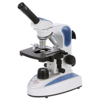
Microscope images with labels. Simple Microscope - Parts, Functions, Diagram and Labelling What is good about transmission electron microscope is that it provides a high degree of magnification and resolution. It is useful in various fields of sciences such as physical and biological science, nanotechnology, metallurgy, and forensic analysis. (1, 2, 3, and 4) Picture 1: The image above is a stereo microscope. PDF Label parts of the Microscope Label parts of the Microscope: . Created Date: 20150715115425Z Explanation and Labelled Images - New York Microscope Company The samples are labeled with fluorophore where they absorb the high-intensity light from the source and emit a lower energy light of longer wavelength. The resulting fluorescent light is then separated from the surrounding radiation with filters, allowing the observer to see only the fluorescing material. Amazing 27 Things Under The Microscope With Diagrams - Microbe Notes Figure: Hair under the microscope. Image Source: Microscope World. Observation under the stereo microscope. Stereo microscopes allow up to 90X magnification for the observation of the general structure and condition of the hair. The external characteristics like color, shape, texture, and length of hair can be seen easily through a ...
Microscope Parts and Functions Eyepiece: The lens the viewer looks through to see the specimen. The eyepiece usually contains a 10X or 15X power lens. Diopter Adjustment: Useful as a means to change focus on one eyepiece so as to correct for any difference in vision between your two eyes. Body tube (Head): The body tube connects the eyepiece to the objective lenses. Arm: The arm connects the body tube to the base of the ... 19,083 Microscope Drawing Images, Stock Photos & Vectors - Shutterstock 19,083 microscope drawing stock photos, vectors, and illustrations are available royalty-free. See microscope drawing stock video clips Image type Orientation People Artists Sort by Popular Science College and University Abstract Designs and Shapes Printing, Typography, and Calligraphy Healthcare and Medical microscope chemistry laboratory biology Parts of a microscope with functions and labeled diagram - Microbe Notes Parts of a microscope with functions and labeled diagram September 17, 2022 by Faith Mokobi Having been constructed in the 16th Century, Microscopes have revolutionalized science with their ability to magnify small objects such as microbial cells, producing images with definitive structures that are identifiable and characterizable. Cell Size and Scale - University of Utah Smaller cells are easily visible under a light microscope. It's even possible to make out structures within the cell, such as the nucleus, mitochondria and chloroplasts. Light microscopes use a system of lenses to magnify an image. The power of a light microscope is limited by the wavelength of visible light, which is about 500 nm.
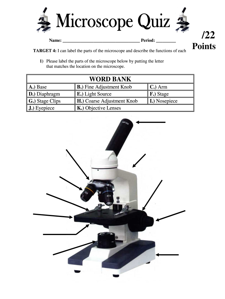
Labeling the Parts of the Microscope | Microscope World Resources Labeling the Parts of the Microscope This activity has been designed for use in homes and schools. Each microscope layout (both blank and the version with answers) are available as PDF downloads. You can view a more in-depth review of each part of the microscope here. Download the Label the Parts of the Microscope PDF printable version here. Word to HTML - Online Converter and Cleaner Free online Word to HTML converter with code cleaning features and easy switch between the visual and source editors. It works perfectly for any document conversion, like Microsoft Word Compound Microscope Parts - Labeled Diagram and their Functions The eyepiece (or ocular lens) is the lens part at the top of a microscope that the viewer looks through. The standard eyepiece has a magnification of 10x. You may exchange with an optional eyepiece ranging from 5x - 30x. [In this figure] The structure inside an eyepiece. The current design of the eyepiece is no longer a single convex lens. Compound Microscope - Diagram (Parts labelled), Principle and Uses See: Labeled Diagram showing differences between compound and simple microscope parts Structural Components The three structural components include 1. Head This is the upper part of the microscope that houses the optical parts 2. Arm This part connects the head with the base and provides stability to the microscope.
Compound Light Microscope Labeling Diagram | Quizlet
Polarizing Microscope Image Gallery | Science Lab - Leica Microsystems The position of the optical axis can be clearly determined with circular polarization. Right: Conoscopic image of the same calcite sample with linear polarized light. The calcite section is perpendicular to the optical axis. Images recorded with a DM4 P microscope using transmitted light, conoscopy, 63x N Plan objective, and polarizers.
2. DATA Sheet FOR Experiment- Microscopy AND THE Cells - DATA ...
Amazon.com: Avery Self-Adhesive Removable Labels, 0.75 x 1 ... Apr 18, 2005 · Marking the expiration dates on white background labels like these are great. We purchased two sizes of the small Avery Labels and they have performed very well. Avery Removable Rectangular Labels, 0.5 x 0.75 Inches, White, Pack of 525 (6737) These are small size labels that we use for marking various products with expiration dates.

This is a common compound microscope. Label its parts from A ...
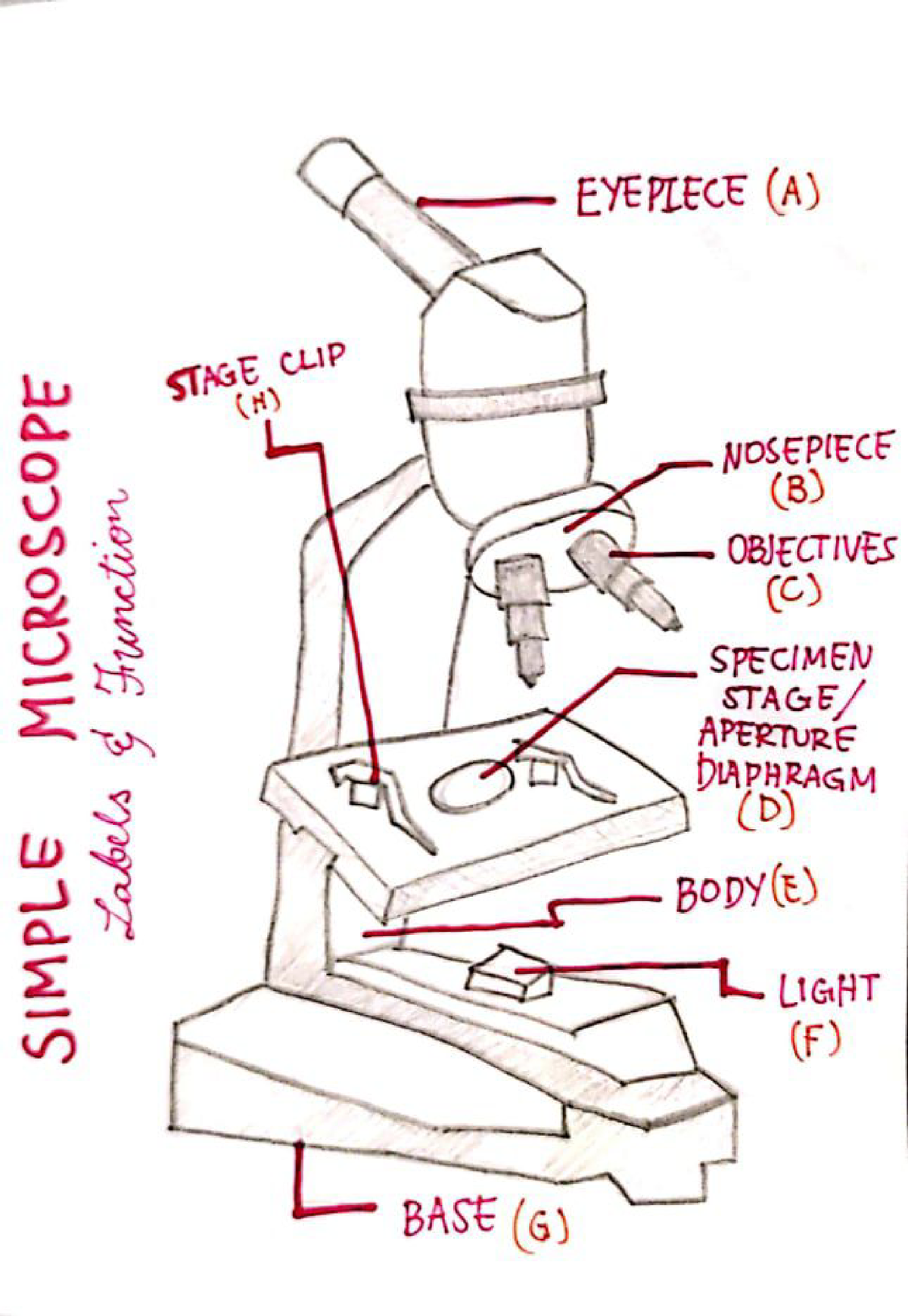
Microscope Types (with labeled diagrams) and Functions The working principle of a simple microscope is that when a lens is held close to the eye, a virtual, magnified and erect image of a specimen is formed at the least possible distance from which a human eye can discern objects clearly. Simple microscope labeled diagram Simple microscope functions It is used in industrial applications like:

Meiji MT6500 Series PCM NIOSH 7400 Asbestos Microscope
Microscope Labeling Game - PurposeGames.com An unregistered player played the game 1 hour ago About this Quiz This is an online quiz called Microscope Labeling Game There is a printable worksheet available for download here so you can take the quiz with pen and paper. This quiz has tags. Click on the tags below to find other quizzes on the same subject. Science microsope Your Skills & Rank
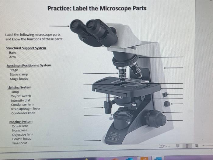
Solved Practice: Label the Microscope Parts Label the | Chegg.com
National Geographic Dual LED Student Microscope Aug 07, 2017 · Buy NATIONAL GEOGRAPHIC Dual LED Student Microscope - 50+ pc Science Kit with 10 Prepared Biological & 10 Blank Slides, Lab Shrimp Experiment, Perfect for School Laboratory, Homeschool & Home Education: Microscopes - Amazon.com FREE DELIVERY possible on eligible purchases

Parts of Stereo Microscope (Dissecting microscope) – labeled ...
400+ Free Microscope & Bacteria Images - Pixabay 413 Free images of Microscope Related Images: bacteria science laboratory research scientist biology lab chemistry microbiology Find your perfect microscope image. Free pictures to download and use in your next project.
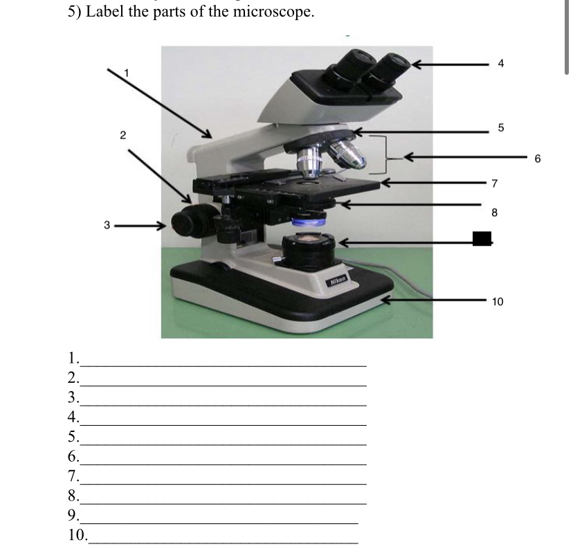
Answered: 5) Label the parts of the microscope. 1… | bartleby
Microscope Stock Photos, Pictures & Royalty-Free Images - iStock the icons include a scientists, laboratory workers, test tube, laboratory scientist using a magnifying glass, petri dish, scientific education, microscope, hourglass, lab worker holding up a beaker, laboratory gown, scientist using a microscope, light bulb, clipboard with checklist, beaker with liquid, laboratory goggles, dna strand, team of …

Dissecting Stereo Microscope Parts and Functions
19,225 Microscope Slide Images, Stock Photos & Vectors - Shutterstock Find Microscope slide stock images in HD and millions of other royalty-free stock photos, illustrations and vectors in the Shutterstock collection. Thousands of new, high-quality pictures added every day.

Biology Lab Microscope Labeling Diagram | Quizlet
A Study of the Microscope and its Functions With a Labeled Diagram ... A Study of the Microscope and its Functions With a Labeled Diagram To better understand the structure and function of a microscope, we need to take a look at the labeled microscope diagrams of the compound and electron microscope. These diagrams clearly explain the functioning of the microscopes along with their respective parts.

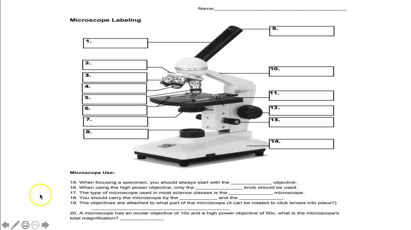
Label a Microscope Worksheet
Skin Histology Slide Identification - Thick and Thin Skin Microscope ... I would like to show you the different histological features from both thick and thin skin histology slides with a labeled diagram. I hope these skin microscope slide labeled diagrams might help you to identify and learn all the structures. If you need more skin microscope slide labeled diagram, please follow anatomy learner on social media. I ...
Microscope- Simple-AND Compound-WITH- Label - BS in Education ...
Electron Microscopy Images - Dartmouth Transmission electron microscope image of a thin section cut through the bronchiolar epithelium of the lung (mouse), which consists of ciliated cells and non-ciliated cells. Image shows the ciliary microtubules in transverse and oblique section. In the cell apex are the basal bodies that are the anchoring sites for the cilia.

Educational / Hobby Microscope (BE211A Eco-Bino-LED)
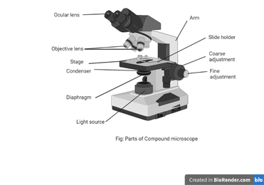
Compound Microscope Parts, Functions, and Labeled Diagram Compound Microscope Definitions for Labels. Eyepiece (ocular lens) with or without Pointer: The part that is looked through at the top of the compound microscope. Eyepieces typically have a magnification between 5x & 30x. Monocular or Binocular Head: Structural support that holds & connects the eyepieces to the objective lenses.

Microscope Diagram Labeled, Unlabeled and Blank | Parts of a ...
Microscope, Microscope Parts, Labeled Diagram, and Functions Microscope, Microscope Parts, Labeled Diagram, and Functions What is Microscope? A microscope is a laboratory instrument used to examine objects that are too small to be seen by the naked eye. It is derived from Ancient Greek words and composed of mikrós, "small" and skopeîn,"to look" or "see".
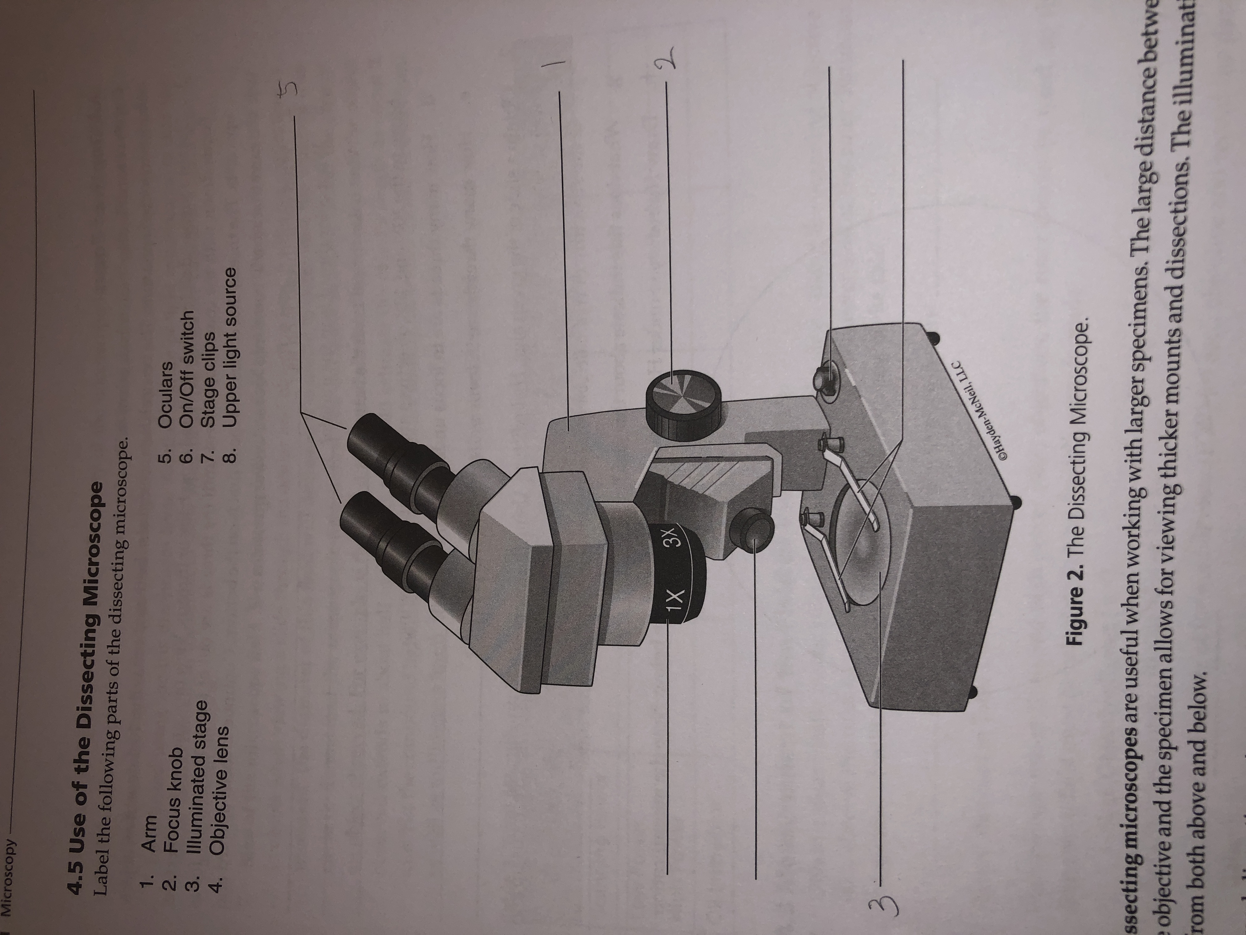
Answered: Microscopy 4.5 Use of the Dissecting… | bartleby
Label the microscope — Science Learning Hub All microscopes share features in common. In this interactive, you can label the different parts of a microscope. Use this with the Microscope parts activity to help students identify and label the main parts of a microscope and then describe their functions. Drag and drop the text labels onto the microscope diagram.

Microscope Labeling Diagram | Quizlet
Microscope illustrations and clipart (70,166) - Can Stock Photo Microscope Illustrations and Stock Art. 70,166 Microscope illustration and vector EPS clipart graphics available to search from thousands of royalty free stock clip art designers. Content Type All Images Photos Illustrations Vectors Video Specific Orientation Primary Color People Search With People Without People Exclude From Results Search Type

Microscope labeling
Microscope Labeling - The Biology Corner The google slides shown below have the same microscope image with the labels for students to copy. I often spend the first day walking students through the steps and having them look at a single slide as we do the steps. Students are often very enthusiastic about using microscopes and will try to start with the high power objective.
Parts of the Microscope Labeling Activity!
Electron microscope - Wikipedia An electron microscope is a microscope that uses a beam of accelerated electrons as a source of illumination. As the wavelength of an electron can be up to 100,000 times shorter than that of visible light photons , electron microscopes have a higher resolving power than light microscopes and can reveal the structure of smaller objects.
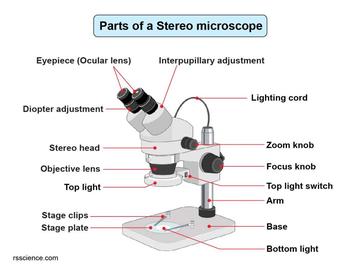
Parts of Stereo Microscope (Dissecting microscope) – labeled ...
Microscope With Labels clip art | Microscope parts, Science diagrams ... Jul 3, 2012 - Download Clker's Microscope With Labels clip art and related images now. Multiple sizes and related images are all free on Clker.com.
Motic B3-220ASC Binocular Microscope Lab Equipment ...
Free Press Release Distribution Service - Pressbox Jun 15, 2019 · Free press release distribution service from Pressbox as well as providing professional copywriting services to targeted audiences globally

Parts of a microscope with functions and labeled diagram
Super-resolution microscopy - Wikipedia Recently, owing to advancements in artificial intelligence computing, deep-learning neural networks have been used for super-resolution enhancing of optical-microscope photographic images, from 40x to 100x, from 20x with an optical microscope to 1500x, comparable to a scanning electron microscope, via a neural lens, and with positron-emission ...

Virtually Labeling a Microscope by Grace Voit | Teachers Pay ...
Parts of the Microscope with Labeling (also Free Printouts) Microscopes are specially created to magnify the image of the subject being studied. This exercise is created to be used in homes and schools. the microscope layout, including the blank and answered versions are available as pdf downloads. Click to Download : Label the Parts of the Microscope (A4) PDF print version.

Label-microscope.docx - Label parts of the Microscope ...
Compound Microscope Parts, Functions, and Labeled Diagram ...
Label microscope - Teaching resources
![750+ Microscope Pictures [HD] | Download Free Images on Unsplash](https://plus.unsplash.com/premium_photo-1661427441367-d0a14fb3f666?ixlib=rb-4.0.3&ixid=MnwxMjA3fDB8MHxzZWFyY2h8MXx8bWljcm9zY29wZXxlbnwwfHwwfHw%3D&w=1000&q=80)
750+ Microscope Pictures [HD] | Download Free Images on Unsplash

Parts of the Microscope with Labeling (also Free Printouts ...

Compound Microscope Parts – Labeled Diagram and their ...
Microscope Labeling
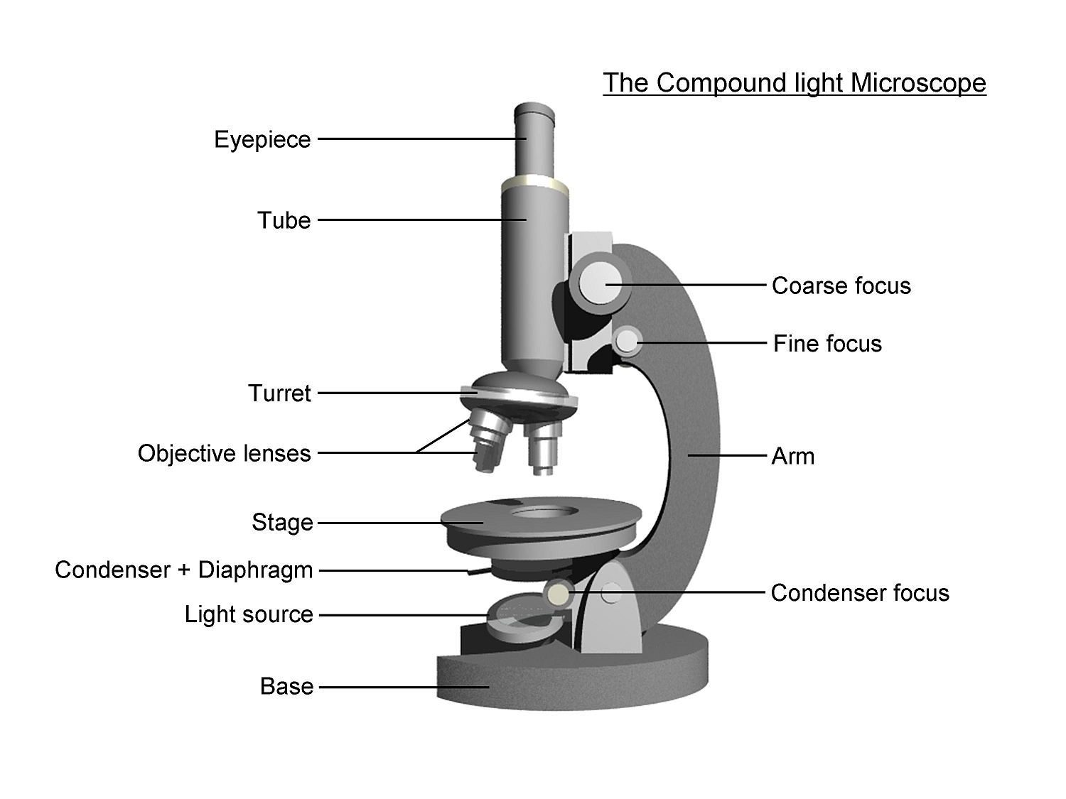
The Compound Light Microscope Label the following parts on ...

Label the microscope — Science Learning Hub

Labeling a Microscope Free Worksheet Pack
Microscope Fill In The Blank - Fill Online, Printable ...
Parts of a Microscope with Their Functions – Microbe Online

Microscope - Label - Part 2 Diagram | Quizlet

Microscope Labeling Part 1 Diagram | Quizlet

Parts of a Microscope Labeling Activity

Label the numbered parts of the microscope - ppt download
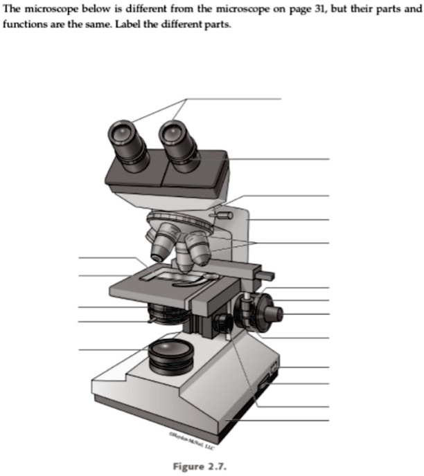
The microscope below i different from the micoscope on Page 31, but their Parts and functions are the sme Label the different Parts, Figure 2.7.

What is a Compound Microscope? | Microscope World Blog
Label microscope - Teaching resources
Label a microscope - Teaching resources

Parts of a Microscope with Their Functions – Microbe Online
Cells and Microscopes

MICROSCOPE Labeling - Part - 3

Collection Of Free Microscopes Drawing Label Clipart ...
Post a Comment for "45 microscope images with labels"